|
|
Esophageal Intramural Pseudodiverticulosis
EIP
General Considerations
- Rare
- Multiple flask-shaped outpouchings in the esophageal wall
- Believed to be a reaction to chronic irritation, such as from
- Reflux
- Alcoholism
- Diabetes mellitus
- Candidiasis
- EIP shows two peak incidences, one in the teens and the other in the 50s and 60s
Clinical Findings
- About 75% have dysphagia
- But it can be found incidentally
Imaging Findings
- Flask-shaped outpouchings into esophageal wall from about 1-4 mm in length and 1-2 mm in width
- They are distributed diffusely in 60% of patients and focally in 40% (upper 14%, middle 14%, and lower 12%)
- Patients often have esophageal strictures, as well, particularly of the upper esophagus
- Intramural tracking is often seen
Differential Diagnosis
- Esophagitis
- Carcinoma of the esophagus
Treatment
- Proton-pump inhibitors
- Dilatation of strictures
Prognosis
- Increased risk of esophageal carcinoma, but no cause and effect shown

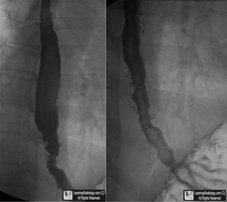
Intramural Pseudodiverticulosis. Two images from a barium esophagram show innumerable,
small outpouchings of barium extending from the lumen into the wall. (white ovals)
There are also linear collections indicating intramural tracking.
For these same photos, click here
For more information, click on the link if you see this icon 
Esophageal intramural pseudodiverticulosis: a reevaluation MS Levine, DN Moolten, H Herlinger and I Laufer.. Am J Roentgenol 1986;147:1165-70
A Case of Esophageal Intramural Pseudodiverticulosis. YE Chon, S Hwang, KS Jung, HJ Lee, SG Lee, SK Shin, and YC Lee. Gut Liver. 2011 March; 5(1): 93–95.
|
|
|
